
Innovation Powers COVID-19 Discoveries
Translational scientists develop, test and implement innovative methods and technologies that make translational research more efficient and effective, thus improving and speeding up the process of translation across many diseases.
Innovation Powers COVID-19 Discoveries
3-D Tissue Models May Speed Virus Drug Testing
Tests used to find drugs with antiviral activity often do not measure a drug’s behavior in the human body. This includes the drug’s ability to stop viruses from infecting cells and the effects of viral mutations on this ability and disease severity. To help address this challenge, our scientists are developing new 3-D tissue models to test how respiratory viruses, like SARS-CoV-2 and influenza viruses, can infect the lungs and other tissues and cause disease. They plan to use these models to predict how well compounds and drugs perform against these and related viruses. Read this February 2021 story to learn more about the role of 3-D models in speeding potential treatments to the clinic.
New Approach for Assessing a Compound’s Activity Could Deepen the Pool of Promising Drug Therapies
Assessing a drug compound by its activity, not simply its structure, is a new approach that could speed up the search for COVID 19 therapies. This action-based focus — called biological activity-based modeling (BABM) — forms the core of a new approach developed by our researchers and others. Our researchers used BABM to figure out dozens of potential anti-SARS-CoV-2 agents. BABM’s impact could extend far beyond the search for COVID 19 treatments to reveal potential therapies for other diseases. Read this March 2021 story to learn more about BABM and its use in COVID 19 research.
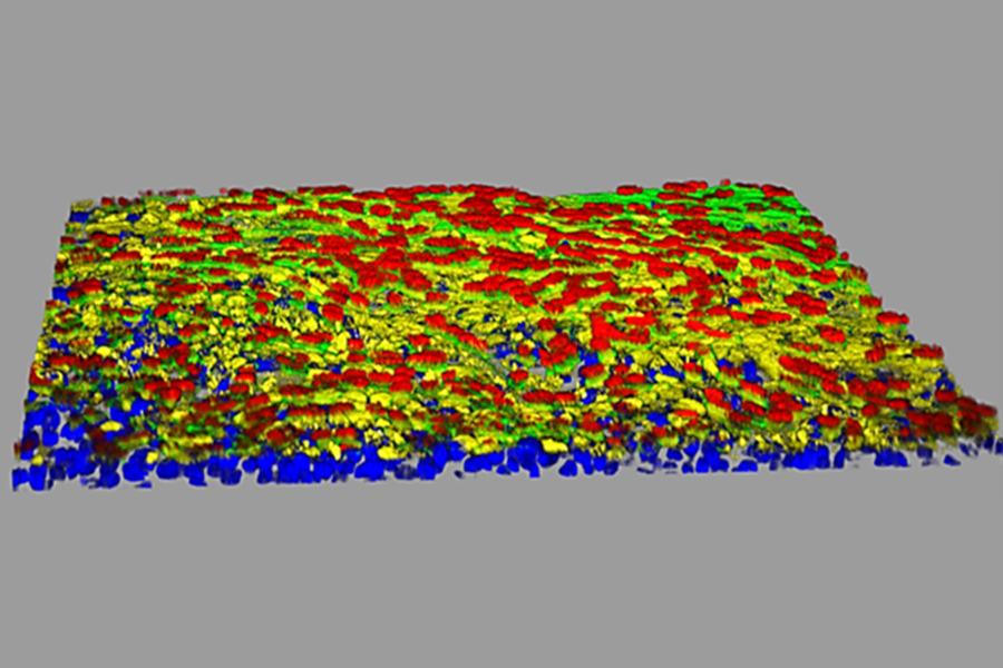
This 3-D model of lung small airway cells is stained with different colored markers that show certain cell types. The yellow represents the receptor protein involved in SARS-CoV-2 infection. (Yen-Ting Tung, Ph.D., and Olive Jung)
Tiny Brain “Organoids” Provide Clues to SARS-CoV-2 Brain Infections
Neurological complications are common in people infected with SARS-CoV-2, but scientists do not fully understand how the virus affects the brain. A team, including our researchers, used brain “organoids,” tiny 3-D tissue models of human brain development, to explore this question. They found that the virus appears to congregate in certain brain cells that constitute a small percentage of overall brain cells. Brain organoids will be a useful tool to better understand how SARS-CoV-2 and other infectious pathogens affect the human brain and to test possible treatments. Read this January 2021 story to learn more about research using brain organoids to study SARS-CoV-2.