NIH-Supported Scientists Uncover Clues to Spinal Cord Development, Neurodegenerative Disease

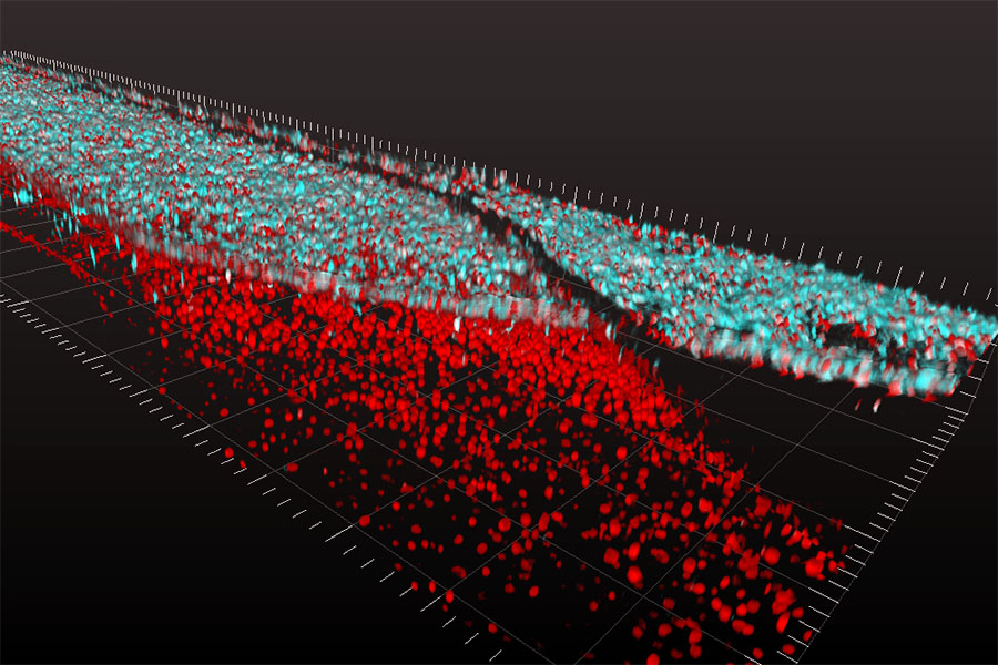
Layers of spinal cord motor nerve cells (top, in blue) and blood vessel cells (bottom, in red) are shown interacting on a tissue chip, a 3-D platform that supports living tissue and cells. (Cedars-Sinai Board of Governors Regenerative Medicine Institute Photo).
Cells in the developing human brain and spinal cord are constantly “talking,” sending chemical signals to help each other grow. But understanding such “crosstalk” isn’t always easy. While scientists have evidence that spinal cord nerve cells, or neurons, interact with blood vessel cells in the brain to help shape the developing spinal cord, there is much yet to be discovered about this process.
To take a closer look, researchers at Cedars-Sinai Medical Center — supported by NCATS and the National Institute of Neurological Disorders and Stroke through the NCATS-led Tissue Chip for Drug Screening program — used stem cells and tissue chips to mimic conditions in the early human spinal cord. Tissue chips are 3-D tissue platforms that model the structure and function of human organs. The results, reported in Stem Cell Reports, could provide a better understanding of the development of some neurodegenerative diseases, such as amyotrophic lateral sclerosis (more commonly known as ALS), and lead to possible treatment strategies.
In this study, investigators first converted adult human skin cells into stem cells, which have the potential to become any type of cell. Then the investigators made these stem cells into either early-stage spinal cord neurons or brain blood vessel cells and grew them together in a specially engineered tissue chip environment that supports living tissues and cells. The researchers found that blood vessels in the brain can trigger the growth and maturation of spinal cord neurons early in development.
“The tissue chip environment provides a better representation of neuron development and how cell types work together than has been possible in the laboratory,” said Cedars-Sinai co-author Clive Svendsen, Ph.D.
Posted May 2018


