
Tissue Chip Researchers Create a Working Model of a Heart Chamber
Scientists can take cells from the skin or blood of adults, turn them back into a type of stem cell called induced pluripotent stem cells (iPSCs) and direct these iPSCs to become heart cells that beat in a dish. But these cells do not have the 3-D structure of the heart, where they line four hollow chambers that contract to pump blood through the body.
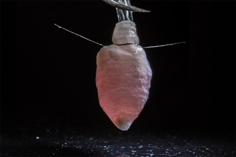
This 3-D model of a human left heart ventricle could be used to study diseases, test drugs and develop new treatments for heart conditions such as arrhythmia. (Harvard University Photo/Luke MacQueen and Michael Rosnach)
Now, researchers supported by NCATS’ Tissue Chip for Drug Screening program have developed a 3-D working model of a human heart chamber. The advance, reported in Nature Biomedical Engineering, is a crucial step toward innovative approaches to study heart diseases, test potential drugs for toxicity or make patient-specific models to test new medications.
The team from Harvard University built a scale model of a left ventricle, the chamber that pumps oxygen-rich blood out to the body. The key was engineering the 3-D ventricle chamber using rat heart cells or human heart cells derived from iPSCs in biodegradable scaffolds. Through ultrasound and other clinical tests, the researchers demonstrated that the model mimics the structure and contractile function of the natural heart.
Using this engineered heart chamber, they developed a model for a type of arrhythmia (irregular heartbeat). This system may provide a way to study arrhythmia heart diseases to understand their causes, to screen drugs for their effects on the heart and to develop new drugs for heart diseases.
Read more about the advance from Harvard University.
Posted December 2018