
3-D Tissue Bioprinting Program Goals
The goal of the 3-D Tissue Bioprinting Program is to speed up the process of discovering and developing new medicines by creating new assay models that better predict the effects of drugs in humans.
About Our 3-D Tissue Bioprinting Program Goals
Contact
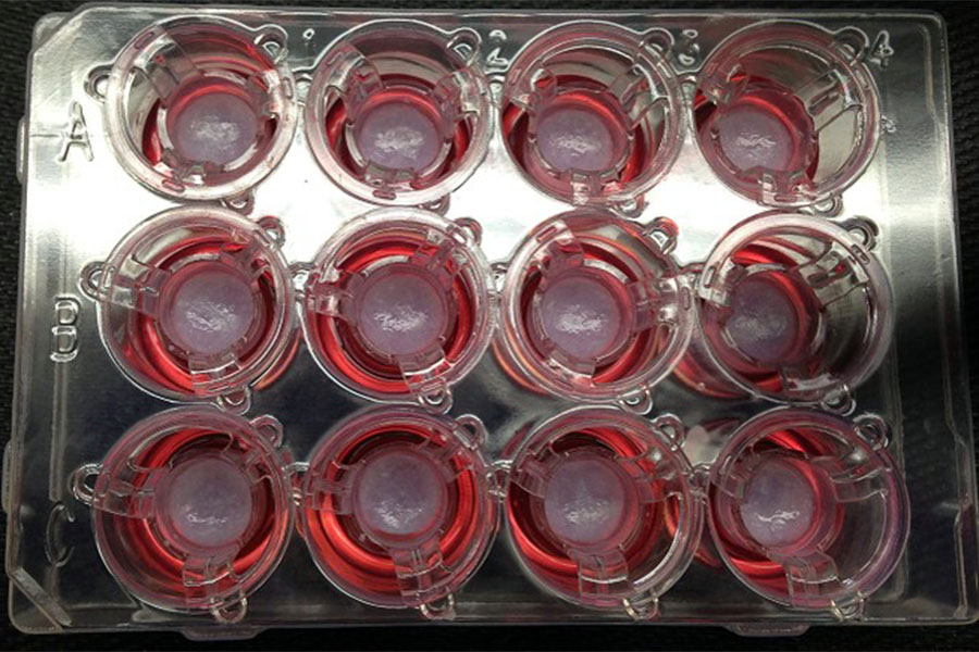
Pictures of 3-D bioprinted tissues in a 12-well transwell plate showing reproducible tissue shape from well to well. (Paige Derr and Kristy Derr, NCATS)
3-D tissue models that mimic characteristics of live human tissues are produced on microplates to test the effectiveness and toxicity of small molecules or other therapeutics. Access to 3-D tissue models in microplate format leverages tissue engineering/organogenesis, stem cell and disease biology, and use of in situ detection technologies (technologies that can be used to detect something in intact tissue) for tissue characterization and testing of drugs’ effects.
The 3-D Tissue Bioprinting Program’s primary focus is developing “disease-relevant tissue models” to reduce the predictability gap between results from current 2-D cell-based assays and results from testing in humans. The program team focuses on many different treatment methods and aims to overcome translational barriers toward the development of urgently needed treatments for unmet medical needs.
While historically the program used bioprinting technologies to make 3-D tissue models, it is now expanding the repertoire of methods used to create 3-D organotypic models for therapeutics discovery and development.
Program Objectives
- Create a portfolio of validated and clinically benchmarked normal and disease 3-D tissue models using a broad range of technologies, including spheroids, organoids, biofabricated tissues and tissue-on-chip, based on a fit-for-purpose approach.
- Apply the use of each 3-D organotypic cellular models as predictive assays in different stages in drug discovery and development, from early discovery to preclinical development.
- Apply biological assay technologies to develop quantitative phenotypic measurements for automated medium throughput screening with 3-D organotypic cellular models.
- Apply laboratory automation and bioengineering solutions to develop and operationalize the use of 3-D organotypic assay platforms for automated medium throughput screening.
- Implement efficacy, toxicology and metabolism screening of small molecules, antibodies, gene therapies and cell-based therapies with 3-D organotypic cellular models in support of therapeutics development projects.
- Operationalize the development and use of 3-D organotypic models using patient-derived cells for advancing precision or "personalized" medicine.
- Operationalize the use of 3-D organotypic cellular models for ex vivo clinical trials to guide clinical trial design, with special emphasis on rare diseases.
- Work in collaboration with the wide biomedical community to advance, demonstrate and disseminate the usefulness and impact of 3-D tissue models in accelerating therapeutics development.
Our vision is to integrate the use of 3-D organotypic cellular models in the drug discovery and development pipelines and effectively implement the principles of the 3Rs (Replacement, Reduction and Refinement) in the humane use of animals in research, and make the process of therapeutic development personalized, faster, more efficient and less costly by decreasing the overall failure rates in clinical trials.