
A New 3-D Skin Tissue Model Helps Identify Possible New Treatments for Herpes Simplex Virus
Jan. 27, 2026
(NCATS)
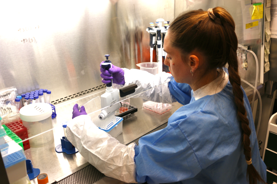
Nearly 90% of potential new drugs fail in clinical trials. This happens because most of these drugs don’t work well in people, even though the results from preclinical studies are promising. To help overcome this issue, NIH launched the 3-D Tissue Bioprinting Program, which applies the techniques of 3-D bioprinting to drug discovery and development. At NCATS, the 3-D Tissue Bioprinting team works with scientists in the biomedical community to make advanced cellular models that mimic human tissues and organs more faithfully.
By using these human 3-D tissue models during drug discovery and development, scientists hope that they will be able to predict better how a drug might work in humans. Marc Ferrer, Ph.D., director of NIH’s 3-D Tissue Bioprinting Laboratory, stated, “By testing the drugs in these assays that better mimic human tissue and organs, we hope that drugs are more likely to be found effective once they move to clinical trials.”
Ferrer highlighted the value of creating a 3-D tissue model in a recent infectious disease study that focused on herpes simplex virus (HSV), published in Nature Communications. HSV is a major public health concern. About two-thirds of people in the world live with the virus, which can cause sores in the mouth and genitals. Having HSV also raises the risk of getting HIV.
Although there is currently no cure for HSV, its symptoms can be managed with acyclovir, the standard of care treatment; however, resistance to acyclovir can develop, particularly in patients with weakened immune systems. As a result, new therapies are needed that can effectively treat HSV infection in all affected individuals.
Ferrer’s team collaborated with Jia Zhu, Ph.D., from the University of Washington to develop a 3-D skin tissue to model HSV infection. Human skin is a layered tissue made mostly of keratinocytes and fibroblasts — the main cell types of the epidermis and dermis, respectively. The epidermis, which is the outermost layer of skin, serves as a protective barrier against environmental threats. The dermis lies beneath it and provides structural support. This collaborative research showed that the bioprinted 3-D skin tissue model closely replicates the structure and layered organization of human skin and that HSV infects these cells the same way it infects people.
Unlike 2-D models, 3-D tissue models include several layers of different cell types, more closely reflecting the structure of human tissues. One key finding of this work was that acyclovir’s ability to suppress HSV infection strongly depends on the type of cells infected. In 2-D cultures, acyclovir was less effective in keratinocytes — the first cells infected by HSV in the skin — than in fibroblasts. The 3-D skin tissue model included the relevant cell types in their proper tissue environment and, therefore, was able to more accurately replicate acyclovir’s efficacy in the skin. In addition, screening 700 compounds using the 3-D skin tissue model identified potent antiviral drugs that might be able to treat HSV infection. These findings highlight the importance of 3-D tissue models for predicting drug responses in cells within a human-like environment that is physiologically accurate. “The hope is that the drug efficacy in these 3-D tissue models will be similar to that in the body, and that we will be able to choose the best drug at the dose that it is most efficacious and least toxic in clinical testing,” stated Ferrer.
Recreating the biological complexity of human skin in this 3-D tissue model is of interest for future studies. HSV can hide in sensory neurons (nerve cells found in the skin), where it waits for stress or other factors to become active again and make symptoms flare up. Developing a 3-D skin tissue model that includes sensory neurons would help scientists understand how the virus “goes to sleep” and find possible treatments for this phase.
Ferrer emphasized that NIH’s 3-D Tissue Bioprinting Program is developing models for many diseases, which expands its impact on patient health. Through the 3-D Tissue Bioprinting Program, NIH is accelerating drug discovery and development for diseases with unmet medical needs.