
NCATS Develops New Technique to Look Inside Cells Growing in 3-D
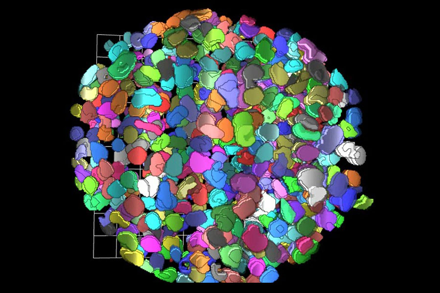
Computed 3-D rendition of a the cells in a tumor sphere model after applying a clearing protocol that enables measurements of fluorescence signal from cell nuclei stained with a fluorescent dye, deep inside the tissues.
Many drugs that seem promising in the laboratory fail during testing in humans. One reason for this failure is that many laboratory tests use cells grown on a flat, two-dimensional (2-D) plastic surface, which is very different from the three-dimensional (3-D) environment in which cells grow in tissue and organs inside the body. A different growth environment might change how cells respond to drugs, and researchers are experimenting with testing drugs in the laboratory using cells that grow in tissue-like 3-D environments.
One technique, which uses cells that have been stained with fluorescent materials that produce light that can be seen with a microscope, can inform researchers about cell behavior or disease. This technique works in laboratory tests when cells are grown in a 2-D environment, where cells are transparent, but it does not work as well in a denser, tissue-like 3-D environment. Current methods to make 3-D tissues transparent and enable cell imaging take too long and are too complicated to be useful for testing thousands of potential new drugs.
Now, in work published in Scientific Reports, NCATS researchers have described a simple, fast process to make 3-D tissues transparent. They imaged tissue with a microscope and saw fluorescent signals coming from cells deep within the tissue. The team developed a computer algorithm that found the nucleus of each cell (where DNA is housed) almost as accurately as a human could — but about 3,000 times faster. The process is automated and eventually could be used to test large numbers of drugs.
The new method enables many types of observations within 3-D growth environments, such as locating molecules inside a cell or finding damaged DNA, and it can do so quickly in thousands of cells in a tissue-like structure.
Posted August 2018