
November 15, 2023: Better Predicting a Medicine’s Safety and Efficacy
NCATS-supported tissue modeling technologies offer a path to more treatments for all people more quickly
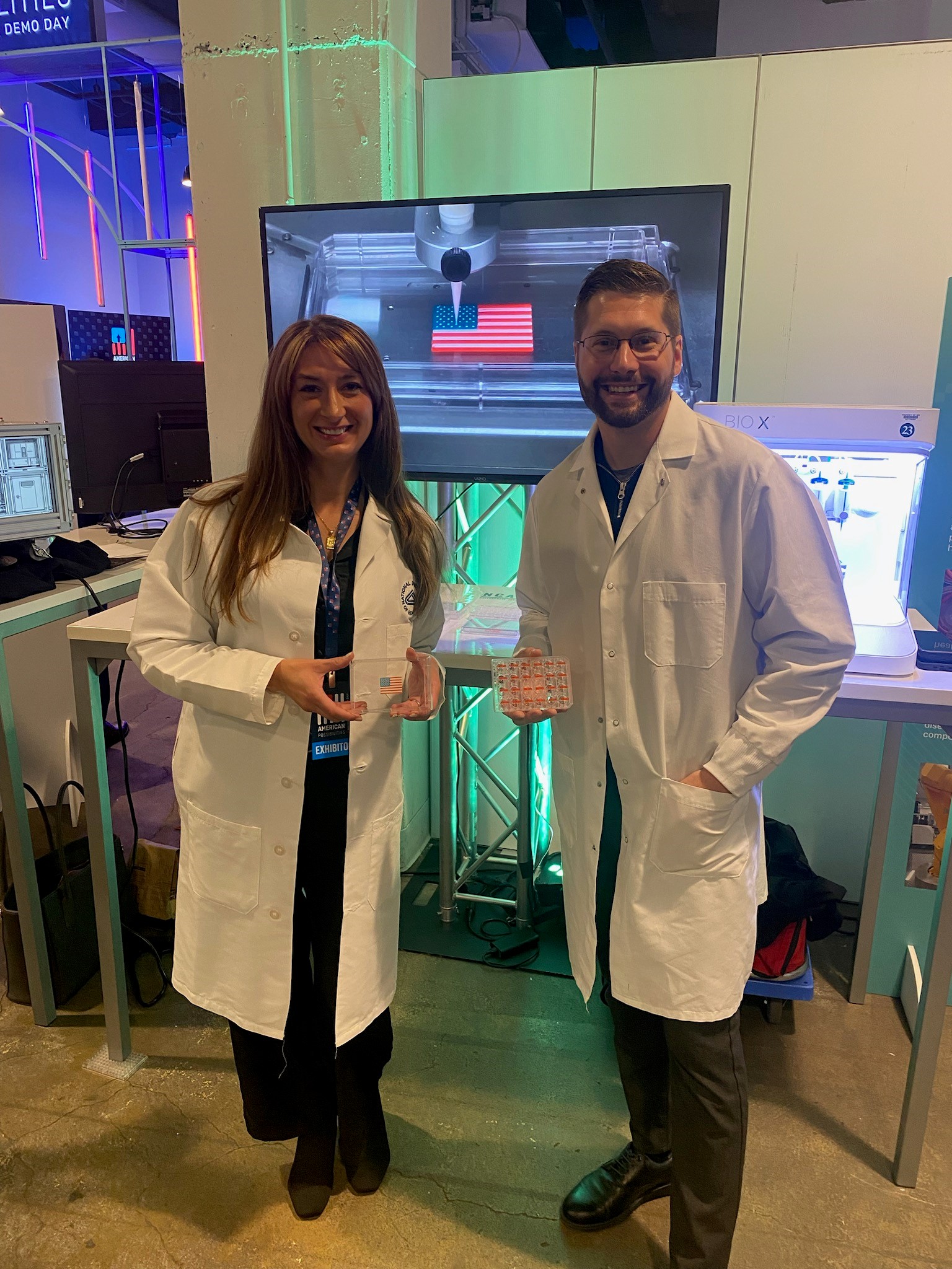
NCATS staff, Kristy Derr and Shayne Frebert, demonstrated our 3-D bioprinting capabilities at American Possibilities: A White House Demo Day on Nov. 7. The event showcased innovations in science and technology made possible by federal investment.
Our vision is more treatments for all people more quickly. A major obstacle we face to achieving that future is the reality that 9 of 10 new medicines that show promise in the laboratory fail in clinical trials because they are either ineffective or too toxic. For many of these medicines, the problem stems from the conventional models used in early testing. As British statistician George Box so aptly put it, “All models are wrong, but some are useful.” The typical animal models and cultured 2-D human cells don’t adequately mimic the structure and function of our cells, tissues, and organs.
Two of our earliest initiatives have tackled this challenge head-on. Our Tissue Chip for Drug Screening and 3-D Tissue Bioprinting programs set out to develop and use 3-D human tissue models for drug discovery and development. Now, with partners and colleagues from around the world, these approaches are getting us closer to the models needed to better predict the safety and efficacy of new medications before they reach clinical trials and, ultimately, save lives.
The evolution of 3-D human tissue models showcases our translational science principles in action, especially the principle of emphasizing creativity and innovation. Tissue chips and 3-D bioprinted models replicate the complexity of our tissues and organs in different ways and even across multiple tissues. Tissue chips capture dynamic processes, like how blood vessels circulate blood to tissue, to study diseases and drugs across organ systems. 3-D bioprinting deposits layers of living human cells and related materials on tiny plates researchers can use to test many potential drug candidates at once. Both types of models can include sensors that allow more precise monitoring of cellular responses. NCATS-supported scientists have developed these 3-D models for nearly every major human organ and tissue system, and the global community of researchers using these approaches has boomed.
Our work with 3-D human tissue models also demonstrates the value of boundary-crossing partnerships. For example, by teaming our tissue chip researchers and space engineers through a partnership with NASA and the International Space Station National Laboratory, we’ve made the technology more compact and easier to use, and we’ve been able to study hard-to-replicate environmental factors, like aging’s effects on cells. The U.S. Food and Drug Administration (FDA) has been an especially vital partner from the very beginning, working with us to consider a pathway for tissue chips as an approved drug development tool. In the bioprinting arena, research collaborations with scientists from NIH, academic institutions, and industry have been key to testing and validating the technology as a solution for both studying and finding treatments for diseases affecting our skin, lungs, eyes, and other organs.
We started to see the direct impact of 3-D human tissue models on the drug development process last year. The FDA based authorization of a drug to treat two rare neuromuscular disorders on data from an NCATS-supported tissue chip. In addition, NCATS-supported researchers used a lung tissue chip to test a potential treatment for inflammatory lung diseases that is now advancing to clinical trials in people. We should see more examples like these soon since the FDA Modernization Act 2.0 clarified that data from preclinical studies not involving animals are acceptable for new drug applications, a required step before clinical trials can start.
We’ve only scratched the surface of how 3-D human tissue models can transform the way we carry out basic, translational, and clinical sciences. In September, we convened experts across fields to explore a way forward for making 3-D cellular models from induced pluripotent stem cells to speed the development of tailored treatments for rare diseases. We also oversee a relatively new program to create tissue chips that include tissues from patients, which could completely redefine clinical trial design and potentially enhance participation in research.
There’s a push for advancing other modeling approaches. We are part of the team organizing a potential new NIH Common Fund program that would advance methods and approaches for more accurately modeling human biology. A related prize competition just launched! We’re also supporting projects to combine the dynamic processes of tissue chips with the high-throughput capabilities of bioprinted models, potentially creating an even more robust modeling platform.
These new modeling approaches will never fully replicate the complexity of our bodies or replace certain animal models that are critical for research; they can, however, provide a powerful complement to them by helping to refine, reduce, or replace depending on the research questions being explored. They offer new possibilities for studying and treating disease in a comprehensive way — one that considers all aspects of health outcomes, including underserved populations, gender, and age.
Now, we can imagine a future of more treatments for you more quickly!
Your partner in science and health,
Joni L. Rutter, Ph.D.
Director
National Center for Advancing Translational Sciences